Analysis
Validation & QC
T/NK Flow Cytometry: COMIP-001, TRBC1 and a 12-Color Panel
A single-tube 13-antibody T/NK flow cytometry panel can make the first diagnostic pass more consistent. Its limits matter: TRBC1 clonality is subset-specific, and published local validation is not universal portability.
What COMIP-001 actually establishes
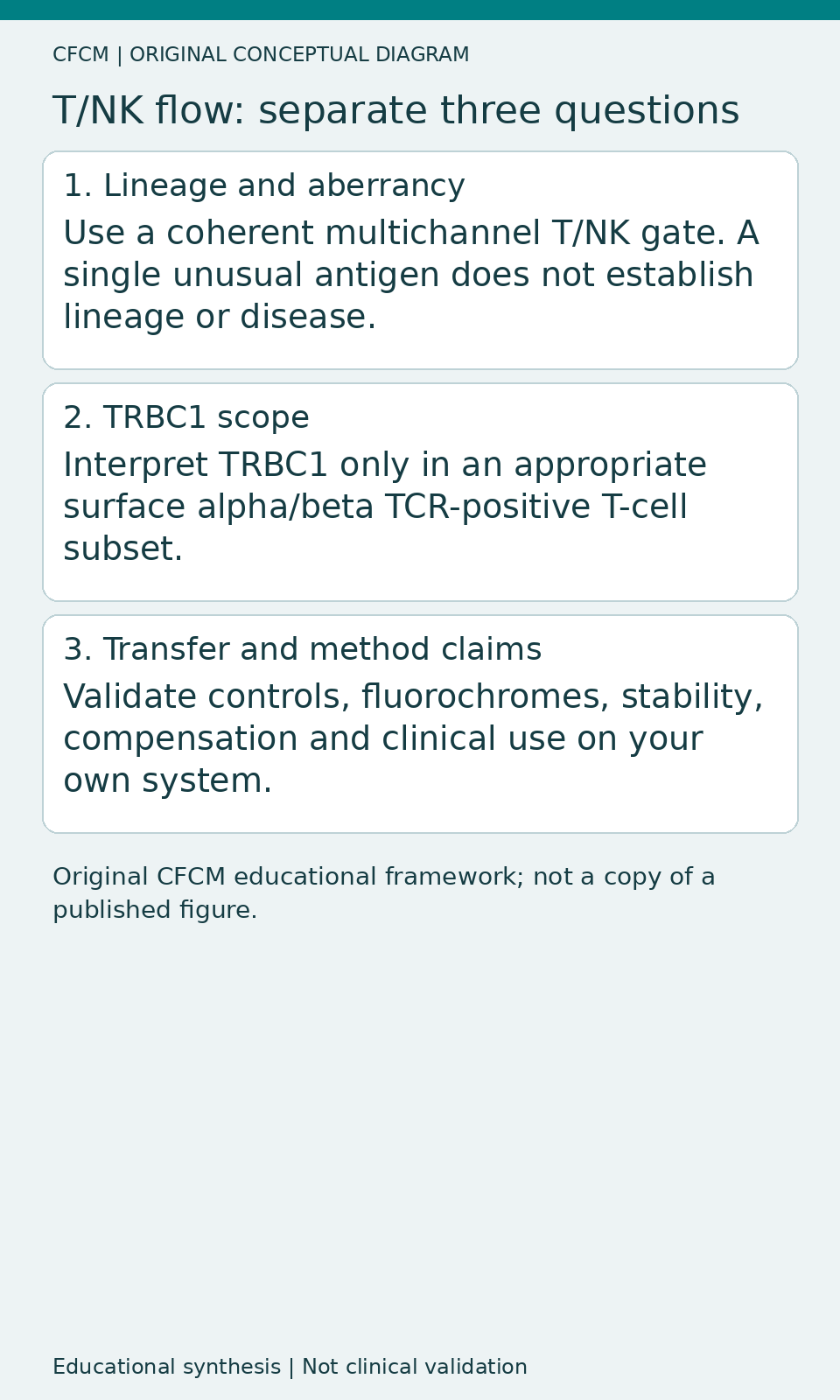
So and colleagues describe COMIP-001, a 13-antibody, 12-color, single-tube immunophenotyping panel for T- and NK-cell leukaemia and lymphoma workups. The authors optimized and validated this panel in one UPMC clinical flow cytometry laboratory using 145 clinical specimens from bone marrow, peripheral blood, tissue and body fluids collected between October 2022 and August 2023. The development is an important attempt at practical panel standardisation, but its local performance cannot be transferred to a different instrument, antibody lot, gating system or case mix by citation alone. The original published report is So et al., Cytometry Part B (2026), DOI 10.1002/cytob.70015.
Why 13 antibodies can fit within 12 colors
Instrument channels and antibodies are not synonyms. A useful diagnostic panel starts with biological questions, then considers fluorochrome brightness, marker co-expression, spreading, controls and the optical configuration. COMIP-001 combines complementary T/NK markers including CD3, CD2, CD5, CD7, CD4, CD8, CD16, CD56, CD57, CD45, TCR-alpha/beta, TCR-gamma/delta and TRBC1. Some combinations share an optical channel in a controlled manner. The purpose is a broad first-pass phenotypic assessment, not unrestricted identification of every rare T-cell entity.
The team selected concentrations by five-point titrations spanning the manufacturer's recommended amount and examined stain index and signal-to-noise ratio. For a local laboratory, this is a reminder that copying clone and fluorochrome labels without checking separation in relevant tissues is not panel transfer. Evaluate the dim antigen-positive fraction, compensation matrix, spread into neighbouring channels, and the effects of abnormal populations before accepting a visually attractive distribution.
TRBC1 is useful, but not a universal T-cell clonality test
TRBC1 assists evaluation of alpha/beta T-cell receptor populations, when the surface receptor complex is sufficiently expressed for an interpretable result. A restricted TRBC1 distribution in a phenotypically defined surface TCR-alpha/beta-positive population can support a clonality hypothesis. It does not demonstrate malignancy on its own: reactive expansions and clinical context still matter. Conversely, a gamma/delta T population or a surface-TCR-negative abnormal population cannot be judged through the same TRBC1 rule. Document the parent population and the number of evaluable events, rather than presenting a TRBC1 percentage without a denominator.
NK-cell abnormalities need a different logic. There is no alpha/beta receptor rearrangement-based clonality readout in NK cells; receptor repertoire, CD56-bright versus CD56-dim composition, CD16, CD57 and tissue-specific reference patterns all affect interpretation. For NK assessments, see the CFCM resource library at cfcm.es and distinguish a phenotypically restricted population from integrated evidence of malignancy.
How strong was the reported validation?
The paper describes clinical concordance, precision, processed specimen and cocktail stability, carryover and a TRBC1 reference interval check using the relevant clinical validation framework. Its 145 specimens came from several specimen types, but specimen diversity is not an external multicentre reproducibility study. The detailed acceptance data reside in an online component not independently evaluated for this CFCM review. Consequently no instrument-agnostic diagnostic sensitivity, interlaboratory agreement percentage or lower limit of detection is asserted here.
In a stability investigation, the antibody cocktail was assessed over eight weeks but reported stable for up to four weeks under the authors' test conditions, while specimens showed acceptable comparability for up to three days. These are two separate stability measurands: an expiry date for a premixed tube is not the same as a shipping window for an incoming specimen. Study-specific conditions do not replace local verification.
The work used a three-laser 12-color BD FACSLyric, FACSuite RUO v1.5 for acquisition, and FCS Express IVD v7 for analysis. An RUO software label in the workflow must not be turned into a regulatory claim about the panel itself; nor does a laboratory validation claim automatically establish IVDR applicability in another jurisdiction.
What it does not do
The authors explicitly exclude cytoplasmic antigen interrogation, measurable residual disease detection, and absolute enumeration from this panel's intended purpose. An AML or B-ALL MRD case cannot be added to the validation scope merely because the cytometer can measure small event fractions. Even within T/NK diagnostics, integrated morphology, clinical history, molecular and histological context remain necessary.
Daily Learning Pill: define the denominator before TRBC1
Ask: in which surface TCR-alpha/beta-positive phenotypic subset was TRBC1 measured, and how many evaluable cells supported that result? If those answers are missing, a neat ratio may provide false reassurance or false suspicion.
Practical next step
For your next T/NK panel comparison, audit clone-fluorochrome pairs, specimen-specific controls, population-specific TRBC1 applicability, and locally justified stability limits. Explore CFCM flow cytometry resources for quality control and validation workflows. This Technical Review is an original editorial synthesis; it is not independent peer review, a clinical guideline or a claim of validation outside the reported laboratory.