Analysis
Validation & QC
Transient Lambda-Expressing Plasma Cells in SFTSV: Restriction Is Not a Diagnosis
A two-centre 2026 SFTSV study reports an infection-associated lambda-skewed circulating plasma-cell expansion. A clinical appraisal separates phenotypic evidence, molecular clonality and malignancy.
A restricted light-chain pattern is a finding, not a final diagnosis
When peripheral-blood plasma cells are markedly enriched for one immunoglobulin light chain, a clinical cytometry service must investigate the possibility of a clonal plasma-cell disorder. But the context of acute infection can produce phenotypes that demand a more cautious interpretation. Tang and colleagues, in a 2026 original research article in Frontiers in Immunology, describe a transient expansion of lambda-expressing plasma cells in severe fever with thrombocytopenia syndrome virus (SFTSV) infection. The critical lesson is not that light-chain restriction becomes unimportant; it is that immunophenotype, time course, clinical setting and evidence of clonality must be evaluated together.
What the study actually examined
The discovery series included 24 adults with SFTS and 20 healthy controls. An independent centre enrolled another 18 infected patients with ten healthy controls. The investigators also compared 67 patients with a mixture of febrile, haematological and inflammatory presentations, and they reported bone-marrow findings in a subset of nine SFTS patients. These are distinct denominators: do not add them together as if all participants underwent one uniform diagnostic accuracy assessment.
Peripheral blood was processed within four hours. Flow methods included absolute lymphocyte enumeration and separate eight-colour immune and plasma-cell phenotyping panels. The reported plasma-cell panel assessed CD38, CD138, CD19, CD20, CD45, CD56 and kappa/lambda light-chain expression. Cytoplasmic light-chain assessment was also described. The work did not prospectively validate a clinical diagnostic threshold for SFTS screening against all relevant alternative diseases.
Lambda enrichment was accompanied by a mature, largely conventional phenotype
The authors found expanded peripheral-blood CD38-high/CD138-positive plasma-cell populations during the acute infection. They reported elevated cytoplasmic lambda expression, especially in severe cases, together with a predominant CD45-positive, CD19-positive, CD20-negative and CD56-negative immunophenotype. Those surface-marker findings differed from the comparison with myeloma-associated abnormal plasma cells in their study.
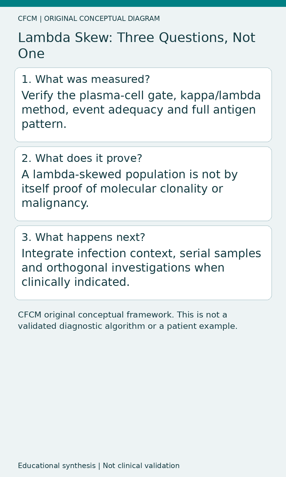
This combination is a warning against equating a striking light-chain distribution with a specific malignancy. It is not proof that every lambda-expressing cell was reactive or polyclonal. The paper explicitly leaves the molecular origin unresolved: an immunoglobulin rearrangement study establishing the precise clonal relationships was not performed. Distinguish a skewed or apparently restricted cytometric population from genetically established clonality and from a clinical diagnosis.
Time course can change the interpretation
The investigators describe a substantial decline of circulating plasma-cell frequency between acute disease and recovery, and a changing lambda-positive fraction during the illness. That temporal pattern differs from assuming a continuously persistent plasma-cell neoplasm. However, a single recovery observation does not independently exclude an underlying haematological condition; if the abnormal population persists or other clinical evidence is suspicious, appropriate haematological work-up remains necessary.
For a laboratory reporting an unexpectedly large plasma-cell population, retain the date of sampling, infection context, treatment history, prior haematology results and whether the same phenotype recurs. Do not retroactively classify an ambiguous specimen as benign simply because the paper describes a transient infection-associated pattern.
Beware of contradictory outcome interpretations
The paper reports associations between lambda-expressing plasma-cell frequencies and measures such as platelet count, lactate dehydrogenase and IL-6. It also compares survivors and non-survivors in small outcome subsets. Such observations are hypothesis-generating. They do not establish a prognostic assay or a universally applicable clinical cut-off. Correlation with severity is not interchangeable with prediction of mortality, and the direction of different comparisons must be checked rather than collapsed into one claim.
What a clinical laboratory should add before using this idea
First, confirm the plasma-cell identity with an appropriate multivariate gate and controls. Second, review kappa and lambda expression using validated cytoplasmic or surface methods as applicable, including denominator adequacy and background. Third, examine CD19, CD45, CD56 and other relevant aberrancies in the whole phenotype. Fourth, interpret the result with clinical, viral and haematological evidence, including follow-up where indicated. Finally, document the uncertainty explicitly: restricted light-chain staining is an observation; the nature and duration of the population are separate questions.
For a formal SFTSV-screening application, a laboratory would need prospective multi-centre recruitment across genuine differential diagnoses, pre-defined cut-offs, adequate event counts, sensitivity and specificity estimates with confidence intervals, and verification of sample handling. The present study does not provide that complete validation. Its comparison cohort did not include some important febrile mimics such as dengue, leptospirosis or anaplasmosis, as acknowledged by the investigators.
Clinical relevance beyond the infection studied
The transferable principle applies whenever a transient immune response can resemble a malignant cell population in one axis of a flow plot. A high-dimensional phenotype and clinical context can prevent premature classification, but no combination of suggestive markers should be called definitively benign or malignant without the appropriate evidence. The clinical question determines which additional tests are warranted.
Daily Learning Pill
Light-chain imbalance, clonality and malignancy are three different claims. Flow cytometry can document the first; proving the second or diagnosing the third may require additional context and orthogonal tests.
What remains uncertain
Mechanism, molecular clonality and external diagnostic performance remain unproven. The independent cohort was small, the observational comparisons were not a prospective screening trial and potential confounding persists. Read the primary methods and discussion before quoting outcome associations. This CFCM educational appraisal uses an original conceptual diagram, not patient dot plots or source figures, and is not independent clinical validation.